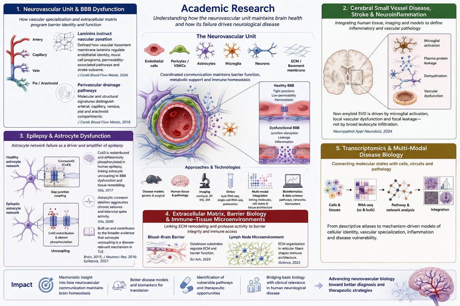
A consistent theme of my work is that disease emerges not only from loss of individual components, but from altered communication between cellular compartments — and that these disruptions can be understood most effectively by integrating functional experiments with molecular profiling.
Translational foundation and vascular pharmacology
At Dr. Reddy's Laboratories (2010–2012), I built my first research foundation in translational pharmacology. I developed statistical and predictive models to estimate the in vivo behavior of candidate formulations, performed pharmacokinetic analyses for preclinical and bioequivalence programs, and worked closely with formulation, analytical, clinical, and regulatory teams. In parallel, my shared first-author review on endothelin receptors in ischemic stroke (Kaundal et al., 2012) reflected an early and enduring interest in vascular mechanisms of neurological disease.
Astrocyte dysfunction in epilepsy
During my PhD at the University of Bonn (2012–2017), I focused on astrocyte dysfunction in temporal lobe epilepsy. Astrocytes form gap-junction-coupled networks that buffer potassium, redistribute metabolites, and help stabilize neuronal activity, so disruption of this syncytium has immediate consequences for circuit excitability.
My major first-author contribution was to explain how this uncoupling occurs. In Deshpande et al. (2017), using human hippocampal tissue from epilepsy surgery together with mouse models, I demonstrated that seizure-associated astrocytic uncoupling is not simply caused by loss of gap junction proteins. Instead, connexin43 was increased overall but redistributed toward perivascular endfeet and showed altered C-terminal phosphorylation at sites relevant to channel permeability. This shifted the mechanistic framework from transcriptional loss to post-translational and trafficking-based dysregulation.
I extended this in Deshpande et al. (2020), showing that constitutive deletion of astrocytic connexins aggravates chronic seizure and interictal spike activity after kainate-induced epilepsy — establishing that intact astrocyte coupling is functionally protective. In parallel, our later work demonstrated that astrocytes in CA1 undergo early cell death during epileptogenesis (Wu et al., 2021).
Neurovascular unit, small vessel disease and vascular zonation
After moving to the University of Münster (2017–2025), I broadened this astrocyte-centered perspective to the full neurovascular unit. In Hannocks et al. (2018), I helped define the molecular architecture of perivascular drainage pathways in the murine brain, showing that pial, arachnoid, arterial, capillary, and venous compartments can be distinguished by specific laminin and structural marker signatures.
In Deshpande et al. (2024), I integrated histopathology, MRI-linked lesion analysis, and mouse–human comparison to show that white matter injury in non-amyloid small vessel disease is characterized by microglial activation, focal plasma protein leakage, and demyelination without substantial peripheral immune-cell infiltration. This refined prevailing models of neuroinflammation and helped identify which mouse models most faithfully recapitulate human disease.
A major culmination of this phase is my recent first-author paper on vascular basement membrane laminins (Deshpande et al., 2026). Using single-cell RNA sequencing, imaging, and functional studies in genetic mouse models, I showed that laminin composition in the vascular basement membrane actively modulates functional zonation across cerebral microvessels. Loss of laminin α4 shifted endothelial identity, altered mural-cell programs, increased permeability-associated pathways, and worsened stroke outcome.
Transcriptomics as a tool, not an endpoint
Across this body of work, transcriptomics has been a central tool rather than an endpoint. I have led or performed bulk and single-cell RNA-seq analyses, reanalyzed public datasets, and built pipelines that link molecular states to imaging and histopathology. This has allowed me to move from descriptive pathology to mechanistic models of how cellular identity is established within anatomical niches and how those states shape neurological disease.